Advancements in medical imaging have revolutionized diagnostic capabilities, allowing healthcare professionals to detect, evaluate, and treat a wide range of conditions with unprecedented accuracy. As clinicians and radiologists seek increasingly detailed information for more precise diagnoses, the demand for cutting-edge imaging technologies continues to rise. For those researching improved imaging techniques, “Microfocus X-Ray Source” often emerges as a key search phrase that promises enhanced resolution and diagnostic confidence.
What Is a Microfocus X Ray Source?
A Microfocus X-ray source allows excellent X-ray beam generation, resulting in high-resolution images that reveal minute anatomical details. Unlike conventional X-ray sources, which emit broader beams, this technology uses a much smaller focal spot—often less than 50 microns—enabling the capture of intricate structures within tissues, bones, and medical devices. This precise targeting is crucial in medical diagnostics, where early detection can significantly impact patient outcomes.
Key Advantages in Medical Diagnostics
The integration of a Microfocus X-ray source into diagnostic workflows offers several notable benefits:
- Exceptional Image Clarity: The fine beam size minimizes geometric unsharpness, resulting in images with sharp, well-defined edges. This enables clinicians to detect micro-fractures, tiny tumors, or subtle abnormalities that may be missed with standard X-ray systems.
- Non-Destructive Evaluation: This technology is especially relevant in fields such as mammography and dental imaging. It enables thorough examination without damaging tissues, making it ideal for repeated assessments and monitoring progress over time.
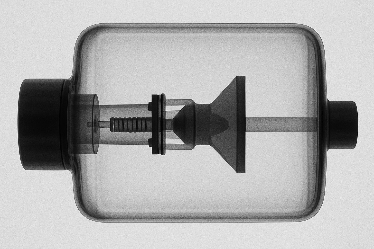
- Enhanced Visualization of Medical Devices: The high resolution provided by a microfocus X-ray source is invaluable for inspecting implants, stents, and other medical devices, ensuring proper placement and identifying potential issues before they become problematic.
Applications Across Medical Fields
The versatility of a Microfocus X Ray Source extends across numerous specialties:
- Orthopedics: Detecting hairline fractures or assessing the integration of orthopedic implants.
- Dentistry: Identifying early-stage cavities and root fractures or evaluating the fit of dental prosthetics with remarkable detail.
- Cardiology: Examining the minute structure of coronary stents and other cardiac devices.
- Oncology: Early detection of microcalcifications and tumors, improving the chances for successful treatment.
By providing such high levels of detail, this technology enables more accurate diagnoses, facilitates better treatment planning, and enhances patient care.
How Microfocus Technology Improves Workflow
In addition to image quality, workflow efficiency is crucial in busy medical settings. Microfocus X-ray systems are designed for rapid image acquisition and processing, reducing patient wait times and increasing throughput. Their precision also means fewer retakes, minimizing radiation exposure, and enhancing patient safety.
Moreover, digital integration capabilities allow seamless sharing and storage of high-resolution images, facilitating collaboration among multidisciplinary teams and supporting telemedicine initiatives.
The Future of Precision Imaging
As medical diagnostics continue to evolve, the adoption of advanced imaging solutions, such as the Microfocus X-ray Source, is poised to become standard practice. Its ability to deliver detailed, reliable images supports early diagnosis, targeted treatment, and continuous patient monitoring—cornerstones of modern healthcare. For healthcare providers seeking a reliable partner in precision imaging, Micro X-Ray Inc. stands out as a trusted solution, offering expertise and innovative technology to advance medical diagnostics and patient outcomes.